Osteoporosis is a disease that wears a progressive systemic nature and is accompanied by a decrease in density indicators with a further change in the structure of bone tissue.
Currently, the issue of the diagnosis of osteoporosis remains fully studied, so the definition of this ailment does not represent any difficulties.

To diagnose patient osteoporosis, comprehensively examine
As a rule, high-quality modern diagnosis of osteoporosis has a complex nature and is based on the evaluation of patient complaints, inspection data, as well as the results of laboratory and instrumental research. On the most informative and common techniques, how to identify osteoporosis, and will be discussed in this article.
Before checking the condition of bones on osteoporosis using laboratory and instrumental methods, it is necessary to determine the factors that could affect the development of the pathological condition of bone tissue.
Make it allows you to carefully collect anamnestic data, examination of the patient and studying its outpatient card.
The most typical factors for the development of osteoporosis are today:
- problems of the gastrointestinal tract, which is accompanied by an impaired calcium suction;
- vitamin D;
- endocrine diseases;
- low body mass coefficient;
- obesity;
- sedentary lifestyle;
- early Climax;
- the presence of spinal deformations and other bone elements;
- insufficient number of calcium-containing products in the food diet;
What should be powered by osteoporosis
- prolonged steroids;
- long recovery period after bone injury.
To learn more about various risk factors for the development of osteoporosis, as well as on its main manifestations and stages, special materials will help, for example, Questionnaire "Osteoporosis in women", "osteoporosis and quality of life", "osteoporosis and pain syndrome".
Early diagnosis of osteoporosis is one of the ways of preventing the development of the disease. About other preventive measures read
Methods of diagnostics osteoporosis
If a person has several risk factors for the development of osteoporosis and burded the bones of the history of the history, the doctor will necessarily offer such a patient to undergo a survey on the definition of bone mineral density, the name of which is densitometry. Analysis on osteoporosis of densitometry, the price of which is completely dependent on the method of its implementation, is an assessment of bone density, that is, the coefficient of their saturation of calcium.
Currently, the most informative methods of diagnosis of osteoporosis is considered:
- ultrasonic computer densitometry;
- x-ray densitometry;
- biochemical blood test for osteoporosis.
More about the method of densitometry you will learn from the video:
Ultrasonic computer densitometry
This is the most common method for diagnosing osteoporosis. The essence of the technique is based on the determination of the speed of passage of ultrasound through the tissue with different density indicators: a high density tissue is much faster than ultrasonic waves, rather than dense structures.
The slower passes ultrasound through the bone, the lower its mineral density, and, consequently, above the degree of osteoporosis.
Ultrasonic survey on osteoporosis is carried out using a special extensive equipment. The doctor, watering the sensor in the projection places of the bones affected by the pathological process, has the ability to display the data obtained to the monitor, as well as write them to digital media in order to study these results in the dynamics. The method of ultrasound densitometry is very sensitive, which allows him to respond with maximum accuracy to the slightest changes in the density of bone tissue.
Such qualities make this method of study effective for the diagnosis of initial forms of the pathological process in the bones, when the loss of mineral density does not exceed 4% of the total.

Ultrasonic computer densitometry - the most common method for diagnosing osteoporosis
The most significant advantages of ultrasound densitometry include:
- the absolute harmlessness of the method, when the analysis of osteoporosis - densitometry with ultrasound waves does not carry any threat to the health and normal life of the human body;
- high research informativeness;
- availability and relatively low cost of the methodology;
- quickness of obtaining results: Ultrasound densitometry Osteoporosis indicators allows you to determine within a few minutes from the start of the study;
- no contraindications to the procedure;
- painless method.
Ultrasonic densitometry does not have contraindications, therefore, it is a universal method of determining the density of bone tissue, which can be used even with respect to people with severe pathologies, pregnant women and children.
Absolute testimony to the study of bones with ultrasound are:
- age (for women it is 40 years old, and for men - 60);
- the first signs of osteoporosis in women, which has become breastfed many times or more than more than a year;
- early or pathological climax;
- frequent fractures;
- violation of the function of the parachitoid glands;
- reception of drugs that wash out calcium bones.
X-ray densitometry
X-ray densitometry is quite accurate, but, unfortunately, not the safest method of determining the bone density.
X-ray during osteoporosis allows you to examine on this disease such a skeleton departments such as a loin, a neck of the hip, a loyal area, a ray-tunny joint and the like.
The study is a very efficient and accurate method, but has a number of contraindications due to its ability to irradiate fabrics.
That is why the diagnosis of osteoporosis in women in an interesting position, children, seriously ill is impossible.
X-ray densitometry, being one of the first methods of studying the health of bone tissue, in our time continues to be improved and developed. Such a tendency to limit the destructive influence on the human body makes it possible to recommend this procedure to an increasing number of patients. To see osteoporosis on the X-ray picture, the doctor allows the unique ability of X-rays to weaken when passing through bone structures, which makes it possible to assess the specialist of their surface mineral density.

X-ray densitometry is a very accurate method for the diagnosis of osteoporosis
X-ray signs of osteoporosis - a reduced amount of mineral substances relative to the total area of \u200b\u200bbone tissue, which the X-ray beam passed. Accuracy and accessibility, and the main high informativeness of this procedure made it an excellent alternative to more expensive ultrasound densitometry.
Both methods have both their positive and, naturally, the negative sides.
Therefore, the question of the feasibility of using one or another version of the diagnosis of osteoporosis in the patient should be solved by an exclusively attending physician.
This method is to determine the metabolic indicators in the bones, as the best option of the patient's additional examination.
Diagnose osteoporosis can not only according to the results of instrumental research. The laboratory diagnosis of osteoporosis will also assume the development of this disease, which is based on the quantitative determination of the hormone levels of the internal secretion glands (thyroid, parachite, genital) in human blood, as well as concentration of trace elements, which are responsible for building bone tissue (calcium, magnesium, phosphorus) , in the morning urine of the patient. These and other indicators in medical practice are called osteoporosis markers And are weighty factors capable of confirming the presence of a pathological process and determine the nature of its origin.

Laboratory diagnostics of osteoporosis will help the doctor diagnose osteoporosis
What tests should be passed during osteoporosis solves the attending physician, relying on the results of densitometric studies, history of the patient, its complaints and the presence of clinical manifestations of the ailment.
Biochemical diagnosis allows not only to determine the disease in the early stages of its development, but also is a very informative method of controlling the effectiveness of the treatment carried out, which has already been an estimate of its effectiveness or inexpediency.
When examining a patient with osteoporosis, the following laboratory studies are carried out in obligatory:
- determining the level of hormones of the thyroid gland (TSH, T4);
- blood test for sex hormones (for men - testosterone, for women - estrogens);
- quantitative study on ionizing calcium;
- definition of parathgamon titers;
- control of the level of active vitamin D (25-hydroxyvitamin D).
Other types and methods for determining osteoporosis
Method that allows you to determine the foci of osteoporosis, which remain unnoticed even with x-ray and tomography, is scintigraphy. It is based on the use of contrast phosphate technetium. The ability of a contrast agent to penetrate the bone tissue depends on the quality of metabolism and blood flow in the affected area.
For details on the method, see the video:
Zones with high blood supply and metabolism, which happen during fractures, metastasis, infectious processes, hyperparathyroidism, look at the scintiogram as "hot foci".
In some cases, survey results need differential diagnostics, for example, to determine the true nature of the pathological process: the presence of hidden fractures, osteoporosis or metastases.
More about what scintigraphy, osteoporosis or metastases are visualized on the scintigram and which there are alternatives to this study, the patient will better explain his attending physician.
MRI study It is a high-tech, innovative and sussuctive method for diagnosing the state of internal organs and organism systems, including the determination of bone density. The results of such a survey make it possible to assess the morphological changes in the tissues and trace their functionality. MRI allows you to obtain a contrast image of internal organs in any plane without ionizing irradiation and the introduction of chemicals. To determine the mineral density of bones, MRI is extremely rare. This is due to the high cost of the method and his tendency to hyperdiagnosis.

To diagnose osteoporosis, MRI is used extremely rare
Estimate the possible risks of development of osteoporosis of bones will help genetic research. A comprehensive genetic study allows you to determine disorders in genes, which is responsible for the synthesis of vitamin D, collagen, the functionality of receptors to pararathgormon and much more. Naturally, even if the method shows a high tendency of a person to develop osteoporosis, it is not a reason to be upset and start being treated immediately. It is enough will be periodic prophylaxis that allow to avoid in the future to reduce the density of bone tissue.
Comprehensive and full-fledged examination allows us to determine the total coefficient of osteoporosis diagnostics. More comprehensive answers to questions about what analysis on osteoporosis is called the most informative study where you can seek help from an experienced doctor. Do not tighten with the appeal to the doctor!
Densitometry is an informative medical examination, his goal is measurement of mineral density of bone tissueman. The procedure is not invasive, painless, allows you to get information about the content of calcium in the bones of a child or an adult, which will help to reveal in a timely manner at the initial stages.
So passes the procedure.
Densitometry can be carried out in different parts of the musculoskeletal system, but it is most often practiced for the study of the following joints:
- knee joints;
- spine;
- hip joints;
- shoulder joints.
Computer, or complex, densitometry at times more informative than normal blood tests and even X-ray. Consider in more detail the types of densitometry, which is the procedure as it is carried out which results shows.
Tasks and essence of the study
Comprehensive densitometry will help to identify:
- The presence at different stages of the flow.
- Bone density level.
- The number of mineral compounds in human bones in any area of \u200b\u200bthe musculoskeletal system.
- Accurate localization of fractures in the spine, the overall state of the spinal column.
- Clarification of diagnoses by bone diseases.
- Establishing a further forecast for the development of osteoporosis, determining the risks of the hip cervix for a few years ahead.
- Evaluation of the effectiveness of the treatment therapy.

The procedure is performed without anesthesia, it is considered safe, since there is no harmful radiation per person. The research method consists in the effects of ultrasound or X-ray radiation; Data is read by sensors and are transmitted to a computer. Next, the special program determines the level of density of human bones.
Computer densitometry is an accurate information technique for detecting osteoporosis at the initial stages. The effects of the rays can determine even minor deviations in bone structures (there is an opportunity to identify even 2% of calcium loss, which indicates the high accuracy of the study).
How to research
How is the densitometry? The research technique depends on the specific type of research, the diagnosed portion of the human body.
General Procedure:
- The patient takes the necessary position on a special table (it is indicated by a doctor, depending on the sector under study).
- If the hip joints are examined, then the person's feet are stacked in a curly bracket.
- You need to lie still. Depending on the method of densitometry used, the duration of the procedure can be from ten minutes before half an hour.
- During the diagnosis, the doctor may ask the patient to delay their breath.
- X-ray beam during the procedure can pass 3 points of bone.
How often can this procedure be done? This is determined individually, based on the overall health and predisposition to bone diseases.
X-ray variety
Two types of densitometry practiced:
- ultrasound procedure;
- x-ray examination.
The ultrasound method is a survey without using rays. Due to the complete safety of the procedure, this type of densitometry is allowed to frequent use even pregnant women and mothers during lactation.
This study is practiced using a special densitometer, which can measure the rate of passage of ultrasound of human bones. The indicator is removed by the sensors and processed in the computer program.
Most often ultrasound examine the heel bone.

Advantages of the ultrasound type of diagnostics:
- Duration - no more than fifteen minutes.
- Lack of harmful radiation or other negative impact on the body.
- Availability.
- Accuracy of the diagnostic procedure.
- Special preparation is not needed.
- The ability to conduct a study, both for primary diagnostics and to control already conducting therapeutic therapy and evaluating its effectiveness.
If the doctor failed to obtain sufficient information from the ultrasound examination of the bones, X-ray densitometry is carried out.

A more accurate diagnostic method - X-ray densitometry. During the procedure, X-rays are sent to the bone tissue of a person. They calculate the amount of mineral substances in bone tissue to determine its density.
Rays of X-rays can identify even minor deviations in the bones. In denunometry, there is much less radiation, rather than in a conventional x-ray, therefore the negative impact on the body is minimal.

Most often, x-ray is used to examine the density of the spinal bones, wrist and hip joint. Also, such a procedure can be carried out for the rest of the areas of the human musculoskeletal system.
Due to the fact that such a kind of densitometry still has radiation from X-ray, it is not recommended to conduct it too often.
It is impossible to say for sure that it is better: ultrasound or x-ray densitometry, since both types of procedures have its pros and cons. However, the more information method is considered to be the study of bones using X-ray.
Where can I do a survey?
You can go through densitometry in the medical diagnostic center. Special attention should be paid not only to the clinic, but also on the qualification of the operator: the quality of the results of the results will depend on it.
The best clinics for performing such a survey:
- Invitro.
- Family doctor.
- Medsi.
- Paterno clinic.
The result of densitometry
A person who is being examined for the first time, it is necessary to understand that it shows densitometry, which bone density standards are distinguished by doctors. The main indicators of densitometry:
- "T" - This is the tissue density indicator compared to the norm. Normal indicator for young people - 1 point and above.
- "Z" - This is the density of the tissue, depending on the age group, to which the patient belongs.
For adult and child, doctors use different scales to assess the results of tissue density.
Deciphering the results obtained in the following table:
With the results of the study, you need to refer to a rheumatologist who will select the course of therapeutic therapy depending on the readings and starting the status.
Traditional coating scheme of osteoporosis:
- : Alostin, Verpein and derivatives.
- Drugs to slow down the loss of bone mass: bonephos, xidone.
- Means for stimulating bone formation (osteogenone).
- Purpose is practiced with pronounced osteoporosis.
- Preparations with calcium,: elevit, complivitis.
With a fracture of the bone, fixation of the limb can be made using gypsum. In more launched cases, the patient requires operational intervention.
Indications for passage
The main indications of densitometry are the following states:
- . It is important to conduct a study of bones at an early stage of this state.
- In preventive purposes The study is carried out by women older than 40 years. As for men, they annually hold such a procedure preferably after 60 years.
- Availability of injuries or fractures Bones in history. It is especially important to diagnose bone density at a spinal fracture or hip joints, since they are most often destroyed under the influence of osteoporosis.
- The presence of severe diseases of the thyroid gland and hormonal failures.
- Women who moved the removal of ovarian (They have increased the risk of osteoporosis).
- Patients close to whose relatives suffered from osteoporosis.
- People who have long taken medicines affecting the washout of calcium from the bones.
- Persons suffering from long alcoholism, smokers with experience.
- People with poorly balanced diet, with a lack of useful substances and calcium.
- Men and women of low height with low body weight.
- Patients practicing fasting for medicinal purposes or for weight loss.
- People leading a sedentary lifestyle.
- Patients regularly providing excessive physical exertion on the body.
Additional readings for densitometry:
- diseases of the spine (, varying degrees of nestness, etc.);
- metabolic disease;
- increased bone fragility;
- indefinite etiology;
- calcium exchange violation;
- severe endocrine diseases;
- general control of the effectiveness of the treatment of therapy under osteoporosis;
- long-term treatment with psychotropic drugs or hormonal contraceptives;
- pregnancy planning period;
- obesity;
- people who often drink coffee.
Contraindications
The ultrasonic type of densitometry is considered safe for humans, so it does not have significant contraindications. As for the X-ray study, due to the radiation impact, it cannot be performed by women during the period of having a child, moms during lactation. If the patient has severe chronic diseases, then before the study, it must necessarily report this to the doctor.
Analysis of bones
Densitometry of bone tissue (ultrasonic, computer) is prescribed by a rheumatologist, however, given the state of a person, the following specialists can recommend the procedure:
- Endocrinologist.
- Gynecologist.
- Orthopedist.
- Surgeon.
If the diagnosis of the state of bone tissue is prescribed an endocrineologist or a gynecologist, it means that a specialist wants to make sure the root cause of the disease and the availability of complications.

Find out what densitometry shows (what it is in general), as it is held, it is possible at a specialist who performs such a study. He will make recommendations on how to prepare for densitometry.
About how densitometry passes, as it is done to diagnose the state of different joints, you can ask the rheumatologist.
Preparation for the procedure
Features of patient preparation for bone examination:
- If the main task of the survey is the diagnosis of osteoporosis, a few days before the procedure, it is necessary to stop taking calcium in any doses and other drugs to strengthen bones.
- Before studying the patient, it is desirable to take off all the decorations, make sure that there are no metal objects (buttons, lightning, etc.) on clothes.
- If a woman is pregnant, then it is important to report this to the doctor before the procedure. It is necessary to make sure that a person has no other contraindications to the study.
- If earlier the patient was already carried out x-ray with the use of a contrast agent, it is important to prevent the diagnostics about this.
Bone density
Some patients fear the negative impact of such a survey. However, the density of bone tissue during densitometry does not suffer, because the procedure does not have a devastating effect as a joint joint.

How often can Densitometry do? Doctors advise to conduct an osteoporosis examination twice a year to people who are in an increased risk area.
As for the prevention of joint pathologies, it is desirable to estimate the overall bone density of bones. Extraordinary conduct of densitometry can be appointed according to the indications (, deterioration of the joints of the joints, etc.). Before such a procedure, it is important to pre-consult with the doctor.
Diagnostics of the spine
The study of the spine and its lumbar department in suspected is performed in the presence of hernia, osteochondrosis or previously suffered vertebral fractures.
X-ray densitometry was shown twice a year with inflammatory pathologies in the spine, scoliosis, large joints (for example, when).
Diagnosis with osteoporosis
The survey on the determination of the density of the bones will allow you to study the composition of bone tissue. Indicators for osteoporosis ("T" and "Z") will be -2.0 and lower.

If the research on osteoporosis reveals this disease, then it will be classified from the results of the tests and the conclusion of the doctor.
How often can I do a densitometry with the already detected osteoporosis? The frequency of surveys depends on the stage of launching the disease, the speed of its progression.
The price of the survey is determined by its species, a specific clinic, an area of \u200b\u200bexamination.
The cost of the study on average is 3500 rubles. In some clinics, the price can reach up to 6000 rubles. Complete densitometry when: timely identified diseases will reduce the risk of developing dangerous and their complications.
Survey of knee joints
In contrast to the conventional Knee X-ray, the densitometry will provide more detailed information on the state of the bone tissue of this joint. The study will allow you to reveal even in the initiative stage when the patient is active until it is observed. This will give a doctor the opportunity to choose a course of treatment for the patient and prevent degenerative joint lesions.
Prevention of osteoporosis development
Osteoporosis leads to the thinning of the bones and increasing their fragility, which provokes fractures. Prevent the loss of bone density, you need to adhere to doctors:
- Lead a healthy lifestyle. It should be completely abandoned by using strong alcoholic beverages, smoking, coffee reception, since all this helps to remove calcium and further remove it from the body.
- Adhere to , the diet of which will be rich in calcium, magnesium and phosphorus. Daily in the menu include meat or fish, greens, cereals, liver, yolk yolks and cheeses. Useful for bones Acoslera products: cottage cheese, kefir, cream.
- Take regularly .
- Women in menopause is important estrogen preparations. They will protect against the development of lack of genital hormones and the negative effects of this state.
- Regularly put on your body physical exertionto strengthen bones and preserve their density. But if a person has already developed osteoporosis, then physical activity will not be so effective.
- Saturate the body with vitamin d. It is recommended at least once a year to go to the sunny regions.
- Not allow obesityas well as critically low body weight.
- Timely treat any chronic pathology, especially the diseases of the kidneys, liver, gastrointestinal tract and hormone malfunctions.
- Every year to see the doctor and conduct diagnostic procedures for the preventive assessment of bone density.
- Avoid hard diets.
Densitometry is an effective way to study the mineral structure of bone tissue, allowing to see a picture of a decrease in the density of bones and identify disorders in its structure. This diagnostic technique is used in osteoporosis and other diseases that cause a decrease in the density of bone tissue. The short procedure is absolutely painless, and will not require special training. As a rule, densitometry is carried out on the lumbar spine, on the hip bones, less often - on the forearm, in some cases the entire skeleton may be conducted.
To date, the usual radiographic study is somewhat outdated, it allows you to diagnose only at 25% of the loss of bone mass. Densitometry of the spine allows you to identify the structural change in bone tissue in the range from 1% to 5% of the total bone mass, which makes it possible to diagnose osteoporosis at the earliest stage. Such diagnosis will allow you to appoint timely treatment and reduce the risk of further development of the disease.
Types of densitometry
- X-ray densitometry (two-energy X-ray absorption). This research method provides the most accurate information about the density of bone tissue. The procedure is based on the use of two different X-rays. Dense bone tissue passes fewer rays. Thus, comparing the results of the absorption of the rays, you can identify deviations in the density of bone tissue. The procedure is carried out quickly quickly, and the dose of irradiation does not carry danger to the patient's health.
- Ultrasonic densitometry. The procedure is based on obtaining data on the speed of movement of ultrasonic waves according to the bone layers, as well as at fixing the scattering of the waves in the bone cavities. The technique is absolutely safe and does not occupy a large amount of time, but has a lower measurement accuracy, rather than a radiological method.
- Quantitative. The procedure allows to obtain a three-dimensional image of the structural density of bones, but since the method highly loads the body with radiation load, it is very rarely used.
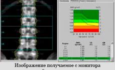
 Nowadays, to diagnose the early stage of osteoporosis, it has become more commonly used ultrasound research methods. This method of diagnosis is an absolutely harmless method, which makes it possible to undergo a survey to children and women during pregnancy. The method allows you to check the various areas of the skeleton with high accuracy. The results of the study are compared with the corresponding norms of indicators, while many patient features are taken into account. The data of the study is displayed on the screen of a densitometer in the form of graphic dependence. The chart is simple enough and does not require special data decryption. The patient immediately receives all the information about the examination, it is diagnosed and prescribed appropriate treatment.
In a situation where ultrasound examination establishes significant rates of bone mass, doctors resort to clarifying diagnostics. To do this, the patient needs to undergo a radiographic densitometry. Radiation irradiation on modern densitometers is very small, and does not harm the health of the patient. This technique will allow not only to establish the exact value of the mineral density of bone tissue, but also to know its strength, elasticity, as well as the thickness of the cortical layer and microstructures.
Passage of diagnosis
Preparation for the procedure
Strict instructions for preparation for densitometry do not exist, but still there are certain points that require attention:
- When using drugs containing calcium, it is necessary to abandon them 24 hours before the diagnosis.
- If you have pacemakers or metal implants, you should inform the doctor in advance.
How is diagnostics?
 You will be asked to lie on the horizontal couch, over which the sensor is located reading information about the degree of absorption of X-rays. The emitter itself is under the couch. In the case of a spinal study, you will be asked to bend legs in hip joints and knees, then put them on the stand. During the diagnostics, the body in a fixed position should be fixed.
Contraindications for radiological densitometry
- Pregnancy or period of breast feeding of a child.
- In the case of CT or with the introduction of a contrast agent for 5 last days.
- When passing radioisotope diagnostics within the last 2 days.
Who needs to be surveyed?
- People predisposed to the development of osteoporosis.
- Women over 45 years old and men over 60 years old.
- Persons over 40 years old who had fractures of various kinds.
- Women who took a long time hormonal drugs.
- People accepting drugs contributing to the washing of calcium from the bones.
- People with endocrine or rheumatic diseases.
- Men and women with insufficient body weight.
- People with osteoporosis identified in the usual x-ray examination.
- People who have various diseases of the spine (, kyphosis,).
- Patients suffering from osteoporosis to appoint effective treatment.
Price on the Densitometry of the Spine
The cost of the densitometry of the spine depends largely on the equipment at which the study is carried out, the method of diagnostics, as well as the authority of the clinic. An examination of one spine will cost approximately 1000-2500 rubles, in most cases a lumbar densitometry is carried out. In the case when the study of the entire skeleton is required, the price may amount to 4000-6000 rubles.
Deciphering the results of densitometry
 In the densitometric apparatus, the denominations of the bone tissue density of the human skeleton are laid, various for each individual plot. Based on these norms, age, gender and individual characteristics of the patient, the analysis of bone indicators is analyzed. The main indicators use:
- BMC (D) is an indicator of mineral bone content.
- BMD (g / cm2) - indicator of mineral bone density.
The results of the study are presented in the form of two main criteria:
- T-criterion - shows the ratio of the bone density in your body to the density of bone tissue completely healthy person of the same sex and age.
- Z-criterion - shows the ratio of bone density in your body to the average bone density indicator of a group of similar sex people and age.
The norm for the T-criterion is the value from "+2" to "-0.9", when the initial stage of osteopying (reducing the density of bone tissue), the numeric data will be located from "-1" to "-2.5". The development of osteoporosis is characterized by the value below "-2.5". In the case of too low z-criteria values, additional research is most often assigned.
Currently, most modern medical centers provide the possibility of conducting a densitometric survey of the spine. Assign the procedure and determine the frequency of its passage should your attending physician.
In the mortality structure from non-infectious pathology, the injury occupies the fourth place, after cardiovascular diseases, oncology and diabetes. With age, the risk of spontaneous fractures increases, which is due to the impairment of mineral substances. The bone decreases the level of calcium, which leads to the development of osteoporosis. In addition, in the people of the elderly complications develop much more often due to the slower fusion of fragments. Early diagnosis and prevention of osteoporosis with densitometry becomes especially relevant.
What is the study and its varieties
Densitometry - (from "Densitas" - density) The method of studying a change in the density of bone tissue, which is used by vertebrologists, traumatologists and therapists for the diagnosis and prevention of osteoporosis.
Osteoporosis is a systemic chronic disease, which is characterized by a decrease in bone mass per unit volume.
The study is based on the determination of the mineral composition of bones, and the percentage ratio of calcium compounds. The most often studied individual peripheral departments (radiation or heating bone, hip joint), which reflect the overall condition of the body.
The procedure is carried out with the help of a special apparatus - a densitometer. Depending on the method of obtaining the result, such types of devices distinguish:
- Ultrasonic - portable monoblock apparatus, which studies the rate of passage of ultrasonic wave through bone tissue. The fabric is denser - the easier it will take place and distinct it.
- X-ray absorptiometry is a method that is used most often. The resulting image is represented as an X-ray image with dedicated zones of different density. There are two types: two-photon absorptionity (the entire skeleton is investigated in 2 or more projections), single-photon (to study the density of a certain area).
- Magnetic resonance imaging.
- CT scan.
The last two methods are rarely used due to their high cost and duration.
Indications and contraindications to conducting densitometry
Talk about osteoporosis as a pathology of people exclusively elderly - a mistake. States that can cause a decrease in the level of calcium in the blood and, accordingly, the density and strength of the bones are many. The study is recommended in such cases:
- Pathology of parathyroid gland: tumors, hypoparathyroidism (the state of reduced functional activity of the gland with a decrease in the secretion of the parathgamon). This hormone contributes to the absorption of calcium from the intestine and reduces its highlighting by the kidneys.
- Fracture of the bone due to a slight injury in history.
- Reception of drugs that reduce calcium levels: steroid hormones (for the treatment of rheumatoid arthritis, bronchial asthma), oral contraceptives, loop diuretics (Furosemid, Toramsemid), anticonvulsant (phenobarbital).
- Alcohol abuse.
- Women over 40 years old and men older than 60.
- People over 30 years old, if the relatives have a diagnosis of osteoporosis.
- Persons who lead a low-wear lifestyle.
- Persons who sit on exhausting diets are subject to significant physical exertion.
- For dynamic observation of the patient's condition and control the effectiveness of treatment.
For women, there is a separate list of indications, since the change in the synthesis of estrogen (female hormones) significantly affects the level of calcium in the blood:
- Women in the period of menopause, especially in the case of its early onset (up to 45 years).
- Women who carried out the operation of admexectomy (ovarian removal may be the step of the extirpation of uterine with appendages).
Densitometry - a gentle procedure, therefore there are no absolute contraindications for her. However, due to the use of X-ray irradiation, the method is contraindicated in pregnant and people who are not able to spend 15 minutes in a horizontal position (spine injury).
How to prepare for densitometry
For the maximum objective results of the study, it is necessary:
- During the day to carry out - to stop the reception of drugs containing calcium, including vitamins (Vitrum, Calcinova, etc.).
- If during the last 2 weeks, a study was conducted with contrasting substances (barium sulfate for intestines, angiography, MRI with contrast) - please inform the doctor.
Important! If there is a probability of pregnancy - the doctor should know about it
In addition, it is recommended to wear comfortable and spacious clothes. The procedure lasts about 15 minutes, during which it is impossible to move. When conducting X-ray densitometry, it is necessary to remove all metal objects, because they affect the result.
How does research passes
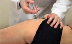
Densitometry is carried out in a specially equipped diagnostic center. The procedure is painless and safe (dose of X-ray irradiation 400 times less than the fluorography obtained).
The patient, depending on the selected method, is located on the table above which the mobile X-ray apparatus is placed or on a couch next to the ultrasound (ultrasonic study).
When X-ray study, the beam goes from the bottom of the table through the area under study and falls on the registering "sleeve", located above the patient. The resulting shot is transmitted to the monitor screen, where analysis is performed using a special computer program. The duration of the procedure is 10-15 minutes.
Ultrasound densitometry implies the use of special sensors that are put on the heel or finger of the patient. Within 3-5 minutes, the passage of ultrasonic wave through bone tissue is carried out.
Advantage of research and possible complications
Before the appearance of densitometry, the only way to diagnose changes in bone tissue was x-ray. However, the informativeness of densitometry is an order of magnitude higher than that of an ordinary X-ray.
The table shows a comparison of the ultrasonic version of the study and radiography.
|
Criterion |
Ultrasound densitometry |
Radiography |
|
Method of obtaining an image |
Bone Ultrasound Wave Movement |
Dispersion of the radiation beam in the tissues of different density with the greatest accumulation in the bones |
|
Visualization of other structures |
Held |
Only bone fabrics |
|
Quantitative: the degree of reduction in bone density is determined |
Qualitative: the presence of changes |
|
|
Diagnosis of osteoporosis |
In the early stages |
In pronounced stages with a loss of at least 30% density |
|
Duration |
||
|
Safety |
Safe study |
X-ray irradiation |
X-ray data allow you to diagnose osteoporosis at the stages when the treatment is ineffective. This method is used to diagnose complications, for example, a vertebral body fracture.
The lack of possible complications after the study is another important advantage of densitometry.
How to decipher the results of densitometry

Analysis of the data obtained during the study is carried out in three main indicators:
- Tissue density expressed in g / cm2.
- T-indicator (hypothetical research statistics). Determined by comparing the resulting density result with a bone density indicator in a woman aged 30 years.
- Z-indicator (standardized). Comparison of the result obtained with the result of a healthy person of the same age and gender.
For the T- and Z indicator there is a single estimate scale, presented in Table:
Doctors recommend to conduct a study every 2 years for people with testimony with the aim of early diagnosis of changes and began preventive treatment of osteoporosis. This method allows you to quickly, painlessly and effectively diagnose changes and prevent possible fractures in the future.
The video presents x-ray two-photon absorptiometry.
An early diagnosis of disease is important for the treatment of bone pathologies. In this regard, the question: Densitometry is carried out, more and more people who have learned about the existence of such a research method. Densitometry of bones makes it possible to identify signs of osteoporosis with sufficient accuracy in early stages, and preventive research using this technology will eliminate the development of the disease in people belonging to the group of increased risk, especially the elderly.
Densitometry is a non-destructive method of determining the mineral density of bone tissue; Basically, based on measuring the level of calcium in bone composition, which largely determines the bone strength. The most important areas of the study are the departments of the spine and the neck of the femoral bone, whose pathologies often lead to the loss of human motor ability. The densitometry of the spine for the elderly, as the level of bone calcium begins to decrease after 35 years and can fall to critical values \u200b\u200bto age 52-55 years.

The main objectives of the study: identifying two main bone pathologies - osteoporosis and osteomalysis. Osteoporosis is characterized by a decrease in tissue density as a result of a decrease in the amount of bone substance in a unit of volume - a quantitative change in bone mass. Osteomalaculation is due to the deficiency of the mineral ingredient under normal volume - a qualitative change in bone composition. If osteoporosis is most often developing against the background of the age degeneration of tissue, then osteomalacia can be observed in pregnant and born women as a result of calcium loss (for example, together with breast milk).
Numerous studies have proven that the problem of osteoporosis is laid in children's age, and the basis of this bone disease is genetic, hormonal, alimentary, mechanical reasons; Chronic diseases and metabolic syndromes. If the children do not form the optimal bone mass, the risk of osteoporosis and bone fractures are imprisoned quite large. Control of the development of a children's bone structure is one of the directions of densitometry.

The risk of osteoporosis is different in different parts of the human skeleton, therefore, when determining the localization of the zone of densitometry, it is important to determine the optimal place of influence. In general, the bone tissue contains two important layers: a compact (cortical) substance, to the greatest degree of bone strength, but having a small speed of metabolic processes, and the spongy (trabecular) substance that is actively involved in metabolism.
Different types of pathology are unenkyly affect these layers. Postmenopausal, hypogonadal and steroid osteoporosis mainly destroy the trabecular substance. The damage to the cortical substance occurs during sedenie, hyperthyroid, hyperparathyroid and diabetic type of disease. If pathology is striking the growing skeleton (under the age of 18), the systemic damage to bones with disorders in both layers is often established.
Currently, many ways to determine the density of bone tissue are known.

The following technologies found practical applications:
- Two-energy X-ray densitometry (absorption) is based on passing two x-rays. The main purpose is the study of the spine and hips. The principle is based on the fact that an increase in tissue density reduces the permeability of such rays. The results of the passage of both rays (on bone and soft tissues) are analyzing and comparison. The accuracy of the method is estimated by about 2% of the change in the mass of the bone per year.
- Peripheral bone densitometry is similar to the previous method, but is designed to study the bones of the limbs, but cannot be used to study the spine and hips. Small portable X-ray sources are used. The accuracy of the method is not sufficiently high, so it is used more often for diagnosis, but to control the process of treating and conducting screening research.
- Two-photon absorptiometry implies radioactive isotopes as sources. Radioactive radiation allows you to determine the density of bone matter in the thigh and vertebrates.
- Quantitative computer tomography is based on obtaining a volumetric pattern of the bone structure when using X-rays. The method is rarely applied due to increased radiation concentration.
- The ultrasound technique is based on determining the rate of propagation of the ultrasonic wave and dispersion in bone tissue. The method is widespread. It has a lower informative ability compared to the X-ray method, but is absolutely safe and accessible.
Densitometry is prescribed by a doctor and it should be passed to people falling into a group of increased risk of bone pathologies, with periodicity of 2 times a year. The risk group is the following people: women aged after 42-45 years old and men older than 55 years; Women in the period of menopause, if he came earlier; women after adxectomy; People after bone fractures; patients with pathology of parathyroid glands; people over 35 years old who have close relatives with osteoporosis; Persons taking medications with calcium flushing effect (glucocorticosteroids, anticoagulants, oral hormonal contraceptives, tranquilizers, diuretic, anticonvulsant and psychotropic drugs); in abuse of alcohol and smoking; people with a fixed way of life and the wrong supply mode; after systemic starvation; people exposed to significant long-term physical overloads; Patients with endocrine and rheumatic diseases, spinal diseases.

It is especially important to conduct research in the appearance of the slightest signs of osteoporosis. Densitometry by radiological methods is contraindicated by pregnant women and nursing mothers. Do not follow the procedure after the introduction of a contrasting composition at previous CT or MRI, if 6-7 days passed. When using radioisotope diagnostics, it is necessary to withstand the appropriate interval. Ultrasonic waves are considered absolutely safe.
Radiological methods of densitometry are carried out using three types of devices: axial X-ray densitometers; Peripheral portable densitometers and peripheral ultrasonometers. Stationary axial devices are used to implement two-energy absocyometry and can perform 4 research programs; Portable - to study the forearm, limbs (especially, heel bone).

This technology allows you to carry out the following studies:
- densitometry of the lumbar spine;
- study of the proximal femur department: the test is subject to the status of the entire department and its parts - the neck (especially, the triangle of Ward), a large spit, a diaphone, anterior zone;
- assessment of the state of the entire skeleton and its parts - spine, pelvis, limbs;
- examination of distal and ultra-dealers of forearm;
- study of the heel bone.
As a result of radiography, the surface mineral density is determined - the number of minerals on the area passed. The study is carried out using fan and point type devices.
Ultrasonic densitometry is based on the use of a portable densimeter. The technique is allowed for examination of pregnant and lactating women. The sensor of the device registers the speed of the passage of the wave depending on the density of the bone. In addition, the broadband weakening of the signal as a result of its absorbing bone tissue is determined.

The results received to the computer, which and analyzes their analysis. Studies are most often carried out in the peripheral areas of the skeleton. The ultrasound densitometry is exposed to the heel and tibia, phalange of the fingers, a patella.
Research criteria
The basis of the densitometric methods of diagnostics is the norms of bone density, which differ in different zones of the skeleton.
Analysis of the data obtained (decoding) is carried out taking into account the established regulatory indicators, gender, age and individual characteristics of a person.
The main indicators of research are considered:
- Navy. The indicator of the mineral composition of bone tissue is measured in grams.
- BMD. Mineral tissue density indicator; Determined in g / cm².
Analysis of indicators is carried out on two main criteria:
- T-criterion. The ratio of the density of the bone density to the density is normal (the value of the indicator for an absolutely healthy person of similar age and gender).
- Z-criterion. The ratio of the resulting density of bone tissue to the average group of people of the same age and gender.
The normal T-criterion is established in the range from -0.9 to +2, which indicates the normal state of bone tissue. When the first signs of bone pathology appears (osteopenia), the T-criterion value is lowered to (-1) - (- 2.5). In the case when the criterion falls below -2.5, we can talk about the development of osteoporosis.
Execution technique and preparation
Preparation for densitometry does not require special procedures. To exclude incorrect effects, only a few limitations are recommended:
- termination of drug reception with calcium additives 24 hours before the study;
- it is necessary to notify the doctor about the following circumstances: passing procedures with the introduction of barium or other contrasting composition; pregnancy; passing examinations with the use of x-ray, tomography or radioactive isotopes; the presence of pacemaker or metal implant;
- compliance with immobility during procedure.
The posture at which the effect of the beam or wave is carried out, depending on which zone of the skeleton is investigated. Above the studied area slowly moves the sensor, the signal from which is projected onto a computer monitor. The duration of the procedure is from 15 to 35 minutes, and for a peripheral survey a few minutes. The resulting result of the study is entered into a radiologist's doctor for subsequent decoding.
With densitometry, the patient remains in a dressed condition, if the clothes are spacious enough and does not have metal elements. Repeated densitometry is recommended to be held with periodicity once every two years to determine the dynamics of changes in bone density.