This is a chronic disease in which degenerative changes of the vertebrae and between the intervertebral disks are between them. Depending on the spine damage site, the osteochondrosis of the cervical, osteochondrosis of the chest and osteochondrosis of the lumbar department. To diagnose the osteochondrosis of the spine, it is necessary to conduct radiography, and in the case of its complications (for example, the hernia of the intervertebral disk) - MRI of the spine. In the treatment of osteochondrosis of the spine, along with medical methods, it is widely used, reflexology, massage, manual therapy, physiotherapy and therapeutic physical culture.
Etiology and pathogenesis
To one degree or another osteochondrosis of the spine develops in all people aged and is one of the processes of aging of the body. Previously, atrophically, atrophic changes arise in the intervertebral disc, however, injuries, diseases and various spinal overloads contribute to the earlier appearance of osteochondrosis. The most often occurs osteochondrosis of the cervical department and osteochondrosis of the lumbar spine.
About 10 osteochondrosis theories are developed: vascular, hormonal, mechanical, hereditary, infectious allergic and others. But none of them gives a complete explanation of the changes occurring in the spine, rather they are complementary by each other.
It is believed that the main point in the occurrence of osteochondrosis is the constant overload of the vertebral motor segment consisting of two adjacent vertebrae with an intervertebral disk located between them. Such an overload may occur as a result of a motor stereotype - posture, individual manner sit and walk. Disorders of the posture, the seat in the wrong position, walking with an uneven vertebral post causes an additional load on discs, ligaments and muscles of the spine. The process can be exacerbated due to the characteristics of the structure of the spine and the insufficiency of the trophic of its tissues caused by hereditary factors. Most often, the defects in the structure are found in the cervical department (kimerly anomaly, cranitonetebral anomalies, anomaly of Kiari) and lead to vascular violations and early appearance of signs of osteochondrosis of the cervical spine.
The occurrence of osteochondrosis of the lumbar department is more often associated with its overload during inclons and lifts of gravity. A healthy intervertebral disc can withstand significant loads due to hydrophilicity located in its center of the pulp nucleus. The kernel contains a large amount of water, and liquids, as you know, have little compressible. The gap of a healthy intervertebral disk can occur at the power of compression of more than 500 kg, while the disk changed as a result of osteochondrosis is broken at a compression of 200 kg. The load of 200 kg is experiencing a lumbar person of the human spine weighing 70 kg, when it holds 15 kilogram load in the tilt position of the body forward 200. Such a large pressure is due to the low value of the pulp nucleus. With increasing inclination to 700, the load on intervertebral discs will be 489 kg. Therefore, often the first clinical manifestations of osteochondrosis of the lumbar spine arise during or after lifting weights, performing work on the house, weeding on a vegetable garden, etc.
With osteochondrosis, the pulp nucleus loses its hydrophilic properties. This is due to disorders in its metabolism or the insufficient intake of the necessary substances. As a result, the intervertebral disk becomes flat and less elastic, radial cracks appear in its fibrous ring when loading. The distance between adjacent vertebrae decreases and they are shifted relative to each other, while the displacement and in the phasetative (arcotted) joints connecting the vertebrae occurs.
The destruction of the connective tissue of the fibrous ring of the disk, ligaments and the capsules of the phasene joints causes the reaction of the immune system and the development of aseptic inflammation with the swelling of the facet joints and their surrounding tissues. Because of the displacement of the vertebral bodies, the capsules of the facet joints occurs, and the modified intervertebral disc is no longer so firmly fixes the bodies of the adjacent vertebrae. The instability of the vertebral segment is formed. Due to instability, the root of the spinal nerve with the development of the root syndrome is possible. With osteochondrosis of the cervical spine, it often occurs during turns of the head, with osteochondrosis of the lumbar department - during the slopes of the body. It is possible to form the functional block of the vertebral motor segment. It is due to a compensatory reduction of vertebral muscles.
The hernia of the intervertebral disk is formed when the disk shifts back, the rear longitudinal bundle is ruptured and the protrusion of the disc part in the spinal channel. If, while in the spinal channel, a pulp nucleus of the disk is squeezed, then such a hernia is called broken. The severity and duration of pain with such hernia is much more than with unexploded. The hernia of the disk may cause root syndrome or spinal cord.
When osteochondrosis, bone expansion occurs with the formation of osteophytes - bone outgrows on bodies and vertebral processes. Osteophytes can also cause spinal cord (compression myelopathy) or cause the development of root syndrome.
Symptoms of osteochondrosis of the spine
The main symptom of the osteochondrosis of the spine is pain. Pain may be acute with high intensity, it is enhanced at the slightest movement in the affected segment and therefore causes the patient to take a forced position. So, with osteochondrosis of the cervical spine, the patient holds his head in the least painful posture and cannot turn it, with an osteochondrosis of the thoracic, the pain is enhanced even with deep breathing, and with osteochondrosis of the lumbar department, the patient is difficult to sit down, get up and walk. Such painful syndrome is characteristic of compression of the root of the spinal nerve.
Approximately in 80% of cases there is a stupid pain of constant character and moderate intensity. In such cases, when inspecting the doctor, it is necessary to differentiate the manifestations of the osteochondrosis of the spine from the myositis of the muscles of the back. A stupid pain in osteochondrosis is due to excessive compensatory tension of the muscles holding the affected vertebral motor segment, inflammatory changes or significant stretching of the intervertebral disk. In patients with such pain, there is no forced position, but the restriction of movements and physical activity is revealed. Patients with osteochondrosis of the cervical spine avoid sharp turns and tons of heads, with osteochondrosis of the lumbar section - slowly sit down and get up, avoid tilts of the body.
All the symptoms of osteochondrosis, manifested only in the area of \u200b\u200bthe spinal column, belong to the vertebral syndrome. All changes that are localized outside the spine are formed extrovelectric syndrome. This may be pain along the peripheral nerves when squeezing their roots at the outlet of the spinal cord. For example, LumboishIngenia is pain along the sedation nerve during osteochondrosis of the lumbar spine. With the osteochondrosis of the cervical spine, this vascular disorders in the vertebro-basilar brain basin caused by the squeezing of the vertebral artery.
Complications of the osteochondrosis of the spine
Complications of osteochondrosis are associated with an intervertebral disk hernia. These include the compression of the spinal cord (discogenous myelopathy), for which it is characterized by numbness, weakness of certain muscle groups of limbs (depending on the level of compression), leading to the appearance of paresis, muscle atrophy, changes in tendon reflexes, urination disorders and defecation. Intervertebral hernia can cause an artery compression that feeds the spinal cord, with the formation of ischemic areas (spinal cord infarction) with the death of nervous cells. This is manifested by the advent of the neurological deficit (violation of movements, loss of sensitivity, trophic disorders), corresponding to the level and prevalence of ischemia.
Diagnosis of osteochondrosis of the spine
The diagnosis of the osteochondrosis of the spine conducts a neurologist or vertebrologist. At the initial stage, the spinal radiography is produced in 2 projections. If necessary, you can shoot a separate spinal segment and shooting in additional projections. For the diagnosis of intervertebral hernia, the assessment of the state of the spinal cord and the detection of complications of osteochondrosis is used magnetic - resonant tomography (MRI of the spine). MRI in the differential diagnosis of osteochondrosis and other diseases of the spine is played a big role: tuberculous spondylitis, osteomyelitis, tumors, behterev's disease, rheumatism, infectious lesions. Sometimes in cases of complicated osteochondrosis of the cervical spine it is necessary to eliminate Siringomyelia. In some cases, if it is impossible to carry out MRI
In medication therapy of osteochondrosis, non-steroidal anti-inflammatory drugs (NSAIDs) are used: diclofenac, nimesulide, lorunoxy, meloxico, ketolalak. With intense pain syndrome, analgesics are shown, for example, analgesic of the central action of flospirtin. Mioryelaxants are used to remove muscle tension - Tolperison, Tizandine. In some cases, it is advisable to appoint anticonvulsant preparations - carbamazepine, gabapentin; Antidepressants, among which preferences give inhibitors of the injecting seizure of serotonin (sertraline, paroxetine).
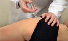
In the occurrence of the root syndrome, the patient is shown inpatient treatment. Perhaps the local administration of glucocorticoids, anti-ease therapy, the use of stretching. In the treatment of osteochondrosis, physiotherapy, reflexotherapy,
Before diagnosing osteochondrosis, it is necessary to conduct a preliminary examination to reveal the ailment. Related signs may be observed with other diseases, so it is very important to find out the cause of osteochondrosis and determine the paths of treatment already at the initial stages.
Osteochondrosis has a number of symptoms, but in addition to them, additional factors should be taken into account. They will help finally make sure the presence of the disease. The diagnosis of osteochondrosis of the cervical and a complex and deep process. It requires the intervention of an experienced specialist, who, as of the patient, will be able to make a diagnosis of accurately.
It is not only not recommended to make self-treatment for self-treatment, but also dangerous for the body. At the very first symptoms, immediately consult a doctor. He will prescribe a deep survey and determines the disease.
Typically, back or lower back pain after any loads quickly hide. If this does not happen, you need to sign up for the reception to the doctor.
- It is worth starting to worry if the head is constantly spinning.
- There is also the upper limbs;
- If you are bothering shivering through the body.
- Discomfort in the chest area, due to breathing difficulties arise.
- Pain in the lower back, passing to gravity in the legs.
- When severe pain in the area of \u200b\u200bthe blades are felt.
- In chronic gastritis
- With pain in the ear or jaw.
These are the exemplary symptoms of osteochondrosis, there are much more of them in reality, and each person, they can manifest themselves specifically. If the patient decided to ask the problem to the doctor, he first generally learns about his condition, but the diagnosis is not in a hurry until a thorough full examination is conducted. It implies a laboratory inspection.
- See also:
How to diagnose cervical osteochondrosis?
Disease recognition includes a number of studies. Before determining the osteochondrosis of the cervical department, the doctor may diagnose in advance, based on the information provided by the patient about his health. After that, he assigns a complete examination of the patient, wringing him a whole list of analyzes that it will have to pass, as well as the mandatory conduct of spinal fluorography. The resulting shot and analyzes will show the exact state of the patient's health. If the doctor is not confident in the diagnosis, it can write additional research. It must be confident that the patient's disorders are in no way related to diseases of blood circulation, hearing and vision or work of the nervous system.

Research methods for the diagnosis of osteochondrosis
X-ray. The radiography method allows you to explore the spine in full. It demonstrates the state of the vertebrae, all violations in the spine immediately can be seen in the pictures, such as highlights, curvature and many others. Also visible between the vertebrae and all the holes are also visible.
In order to recognize osteochondrosis of the breast or cervical system, you need to carry out x-ray twice.
The first time, lying on the side, and the second - in the literal position on the back. Both snapshot are presented to the state of the vertebrae. The doctor may also recommend making a radiot of mouth.
Tomographic method. Based on a magnetic resonance and computer study. This method of diagnosing osteochondrosis can be carried out in two ways. The first is considered the most productive, since thanks to him, the vertebral is clearly visible, they are not observed by the adjacent organs of the body. Also, the snapshot shows vessels and nerves. Such a kind of tomography allows you to determine the presence of many diseases of the spine in the body of many diseases and, in addition, determines the exact location of the defect. The second method shows whether the hernia is used to provide data on any changes in the spine.
- We advise you to read:

Laboratory tests. It is used to compile a complete vision of the disease, the blood and other elements are investigated in this method. The detected osteochondrosis always leads to a change in component parts of the blood. This method allows not only to determine whether there is a disorder in the spine, but also to understand what kind of disease is disturbing the patient and definitely make a diagnosis. Already having analyzes, the doctor decides, continue the survey on or not. It pays special attention to the presence of calcium in the blood and on other nuances.
- We advise you to read:
Differential diagnosis To determine the cervical osteochondrosis has a special place, because it helps to determine which disease is disturbing the patient. The method shows, in a patient osteochondrosis or other disease with similar symptoms.
The procedure helps to understand whether there is a nervous, circulatory and other systems in the body of pathology.
- Be sure to read:
The meaning of the diagnosis in a wide and deep study of the body. The patient must pass such procedures as ultrasound, cardiogram and many others. It is a survey on the presence of heart disease, digestive and other systems. Of all these methods, the longest and expensive, but most effective.
What can be confused osteochondrosis?
There are a number of diseases with similar symptoms. Among them:
- Pathology in the growth of the spine, deformity and defects. This may be after surgery, injury, fall or in the case of the formation of a benign or malignant tumor.
- Berdy Diseases, because of which the spine can sow. Lameness.
- Diseases of internal organ systems that can be seen only conducting a complete study that includes differential diagnosis. This includes many diseases of the urinary system, cardiovascular pathologies and diseases of the digestive system.
- Problems with nerve fibers.
Without bringing research to the end, you can confuse the disease with a similar, thereby defining incorrect treatment. This may lead to negative consequences in the future. Even if the spine ceases to hurt, after a while he can let him know about himself.
| Your feedback on the article |
- the disease is not only dangerous, but also hidden. You can diagnose it in the early stages only by chance (because the symptoms are not manifested), and in the later it is almost irreversible. Treatment in such cases is aimed at mitigating symptoms and prevent further development. But in order to treat the disease, you need to know what to specifically treat, and here the diagnosis of osteochondrosis of the cervical area shows all its variety of methods. The article will describe the most common of them.
On the importance of diagnosis
Early diagnosis of the cervical osteochondrosis can save the patient from constant painful pain, disability and even death.
The reason has already been highlighted above: treatment is aimed at symptoms and affiliated diseases, and not to the sick neck itself. For example, if a patient has a cervical radiculitis due to degenerative-dystrophic changes, it is possible to assign such treatment that will be removed for a while.
But fully prevent relapses if possible very difficult.
Ask your question to a neurologist for free
Irina Martynova. She graduated from Voronezh State Medical University. N.N. Burdenko. Clinical ordinator and neurologist Buz in \\ "Moscow Polyclinic \\".
Therefore, the earlier doctors diagnose the disease, the earlier it stops, and the less the secondary pathologies will arise.
What doctor to contact?
With osteochondrosis of the cervical department in diagnosis and treatment, 4 doctors can participate:
- Therapist. "The first line of defense", a doctor who understands everything. The therapist itself does not engage in osteochondrosis, but on the basis of complaints and differential diagnostics, it can come to a preliminary conclusion and give the direction to a specialist of a narrower profile.
- Surgeon. If you follow logic, the diagnosis must put the surgeon, "specializing" on the spine. The orthopedist surgeon on the basis of X-ray, CT or MRI can very accurately assess the state of the vertebrae and give the necessary recommendations.
- Cardiologist. This doctor does not very often take part in the survey, since its conclusion is necessary only when a large cervical vena or artery appeared by the spine. On the other hand, it is precisely such problems with the most dangerous, so the cardiologist may unexpectedly become a leading physician in treatment.
- Neurologist. Since 99% osteochondrosis is accompanied by a radiculitis (pain due to nerve), neurologists are most often osteochondrosis and are engaged. A good neurologist can all: and make a decision on the need for magnetic resonance imaging, and recognize problems with vessels, and prescribe treatment, and (in acute need) to send a patient for advice with a surgeon about the operation.
Diagnostic methods
Anamnesis
Any diagnosis starts with Anamneza. At this stage, most diseases are diagnosed. In the case of degenerative processes, the doctor should ask about the following things:
- Why did the patient apply for help? At this stage there is a complaints.
- How long have the symptoms manifest? Here is the dynamics.
- Did the patient deal with independent treatment? Depending on the response, the dynamics may have to be corrected.
- Did there be similar signs in the family? Confirm / eliminate heredity.
- Are there any additional symptoms? Information is important for differential diagnostics.
In general, with osteochondrosis, these questions are suitable for both the cervical department and for any other.
Symptoms
 How to determine osteochondrosis of the cervical department? Ask where it hurts.
The symptom survey is crucial. Since pain is manifested almost in all cases and has its own characteristic features, on the basis of some symptoms, you can make a fairly accurate conclusion that will later be confirmed tool. The most important symptoms:
- starting with the vertebrae;
- pain in a strictly defined place:, cheek, nose, tongue, tongue and others;
- episodes of paresthesia (), which arose without visible reasons;
- limited mobility in hand, neck;
- hypertension, fainting;
- , Depression, sleep disorders, other mental disorders.
Inspection
How to diagnose problems with vertebrae? Look at the spine.
The spine is a large structure that lies pretty close to the skin. This makes an inspection of a very effective diagnostic tool. The entire vertebral post falls under the attention, ranging from the waist and ending with the neck. When examining, the doctor reveals such defects as scoliosis, kyphosis and other curvatures (which often become the cause of osteochondrosis).
In particularly launched cases, you can see the displacement of a certain vertebra with a naked eye.
Radiography
 X-ray - the most affordable waywhich can be determined osteochondrosis.
The bottom line is that ionizing radiation passes through the patient's body, and some of this radiation is lost on the road. At the exit, it turns out a picture with inhomogeneous "indexing", reflecting the condition of the internal organs.
Radiography is necessarily done in two planes (from the back and from the side), because one two-dimensional picture cannot display a three-dimensional structure.
pros:
- low price (400-600 rubles);
- wide accessibility.
Minuses:
- bad (compared to CT and MRI) image quality;
- the pictures shows only the external state of the vertebrae;
- radiation is not harmless.
How to identify problems in the picture? With a radiograph, everything is quite simple. Example:

If you look at the bottom cervical vertebrae, we can see that there is no dark area between them, which is between the rest of the vertebrae.
This means that the intervertebral disk felt, and the vertebrae comes into contact with each other.
Another, more informative example with explanations:

Functional radiography
This method is subspecies of ordinary x-ray. Snapshots are made mainly in the lateral projection, and the patient is asked, for example, to push the head back. With this approach, it is possible to get not just a picture with vertebrae, but also to estimate their functionality (hence the name of the method).
Kt.
 Computer tomography is next stage of radiography development.
A special device with high speed makes many snapshots, and then with the help of algorithms restores the information received to a full-fledged 3D image with high quality.
There is such a procedure around 3000-4000 rubles.
pros:
- the quality of the pictures is higher compared to x-ray.
Minuses:
- exposure is higher than 120 times compared with a single snapshot;
- the price is above average.
The resulting pictures can be analyzed independently. For example:

The picture shows that one of the vertebrae is pretty deformed. One more example:

Snapshot - in two projections. It can be seen on the left that bone growths are formed on the vertebral, which indicates spondyliz (complication of osteochondrosis).
On the right one can notice the same growths.
MRI
 Magnetic resonance imaging - the most popular diagnostic method.
The essence lies in the fact that a huge magnet makes certain atoms enter with it in resonance, which is fixed by special devices. The snapshot is obtained very clear and divided into layers (which helps to look inside the organs).
Due to the lack of radiation, the MRI apparatus is quite safe.
pros:
- very high-quality pictures;
- safety.
Minuses:
- price (about 5000 rubles);
- inaccessibility (residents of small cities sometimes have to stand in semi-annual queues or go to other cities).
The uninitiated in the topographic anatomy is quite difficult to read the pictures of MRI alone, but it is still possible to notice something.
 Blood and urine tests don't play a big role In the diagnosis of cervical osteochondrosis, but can be very help with differential diagnosis. Full analysis of tests during diph. Diagnostics will take the volume of the book, so we simply give two indicators that can help in the osteochondrosis diagnosis:
- Daily urine volume. The reduction in daily volume may indicate that the left vertebral or intervertebral hernia closed the artery. The decline in this parameter is accompanied by edema.
- ESO (erythrocyte sedimentation rate). The high level of ESO explicitly indicates an inflammatory process in the body. Soe can rise in exacerbation of the cervical osteochondrosis, accompanied by inflammation.
Other methods
Sometimes ultrasound doppler - procedure at which get a full vessel card.
UDG can show the true cause of headaches, loss of consciousness, psychosis and other symptoms associated with insufficient blood flow.
Look useful videos on this topic
Complexities and nuances of diagnostics
Two main problems of diagnosing degenerative processes in the spine: low speed and high price. Even if the doctor can understand that in front of him is a patient with osteochondrosis, at this moment the diagnostics just begins.
It is necessary to appreciate: how many vertebrae is involved in the process, as far as the intervertebral discs were injured, whether osteophytes did not arise, how launched radiculitis and how large neck vessels feel. All this takes precious time. This time can be won by paid procedures (CT, MRI), but the price is already up. Naturally, the experience of the doctor himself plays a latter role. All this in the amount makes the diagnosis of cervical osteochondrosis with a difficult task for both the doctor and the patient.
The cervical osteochondrosis is a difficult task in which several doctors can be involved. It includes not only standard methods (collection of history and analyzes, differential diagnosis, inspection), but also a plurality of instrumental methods (radiography. Normal and functional, CT, MRI, Doppler). The formulation of an accurate diagnosis may pour out large financial spending, but the faster the treatment will begin, the more damage will be the effects of the disease.
Osteochondrosis of the spine - the scientific name of the disease The degenerative-dystrophic vertebrogenic process. It is degenerative, due to the fact that it is replaced by working fabrics to functionally defective - this is the deposition of calcium salts in cartilage, disks, muscles and the growth of connective tissue in the place of damage. And the dystrophic is due to tissue nutrition. Vetebrogenic - it is emanating from the spine, but affecting and muscles, joints, skin, but in some cases internal organs.
Osteochondrosis: causes and development of osteochondrosis
It is believed that the development of osteochondrosis is associated with the impact on the body of the set of factors. An important factor is daily load on the spine . At the age of twenty years, a decrease in intervertebral discs is already observed, liquid and elasticity is lost, the connection between the vertebrae and the flexibility of the spine itself is disturbed, while the intervertebral discs can no longer receive power from blood flow, and remove it from the surrounding tissues. But mostly osteochondrosis begins to develop after forty years. And at the age of fifty - sixty years, the amount of fluid in the disk-laying is only a little more than half.
Also to illness can lead spine injury , congenital or acquired anomalies anatomical relationship in the spine department. The development of the disease can affect lifestyle, occupation, professional sports. Osteochondrosis Also, many tend to consider the disease not only the spine, but also the entire body associating the mechanisms of development with the immune autoagression of the connective tissue, and sometimes the violation in the vertebral vessel system, leading to the edema in the field of nerve endings.
But among the factors affecting the development of the disease, you can also call inheritance reasons. In recent years, hereditary impaired of the formation of one of the main components of intervertebral disks and ligaments - collagen 2 and 3 types has been revealed. The hereditary impairment of muscles, skin, skeleton is also isolated. The disease is detected in children who have not yet had loads on the spine, but changes in it are already detected.
Painful changes, disk degeneration begins in the kernel when the amount of fluid decreases, and the discs begin to dry. Lose the main shock-absorbing function. Two vertebra and lying inter-disk form a spinal segment. If one of the segments becomes still, changes begin to occur in the entire vertebral post. The closest segments are forced to take on the functions of the affected segment, while the load on them is excessive. Over time, this may not deal with these segments. If the segment loses mobility, its nutrition is immediately disturbed.
When the height of the disk decreases, the pressure on the fibrous ring increases, and that part of which accounts for the greatest load, loses its density and shape, and with time it is deformed, which already leads to pain, and when the disease is developed, the fibrous ring is broken, and the kernel elements, Going beyond the limits shape the hrying of the vertebral disk. These changes cause pain in the affected segment. The clinic of the disease is associated not only with the lesion of intervertebral discs, but already in the early stages there are changes in intervertebral joints as a result of a violation of the compliance of the joint surfaces, almost the articular capsule, disorders and equilibrium in the spinal column. In this case, the bone vertebral tissue reacts to changes in the sealing layer and the formation of osteophytes. As a result, the formation of battles in the spinal channel and the development of its narrowing.
Osteochondrosis and its forms
There is osteochondrosis of the cervical, thoracic and lumbar spine. Osteochondrosis is also found when more than one spine is affected and generacted with total lesion. The most common is cervical and lumbar forms. This is due to great mobility in these spinal departments.
Cervical osteochondrosis - the most common and most difficult form of the disease. Leading symptoms in the clinic of the cervical osteochondrosis are a root, vegetative-dystrophic and spinal. A combination of symptoms can be observed with a predominance of one of them. The main symptom in this form is pain, and its localization depends on the level of the lesion - it may be a clavicle and shoulder, the entire upper limb or the front surface of the chest. Also characteristic of the limitations of the mobility of the cervical department, crunch when turning. When irritating the nerve roots of the cervical plexus, the shoulder pain appears, which is in hand, it is hard to raise it, pain and in a shovel arises. Vegetative-dystrophic syndrome is characterized by sharply in the neck, especially in the subtlety zone. At the same time, the neck muscles are in constant tone. Sometimes pains become minimizing, spreading to the arm, while hard to take the hand to the side, the fingers of the hands may be in motion, the skin is pale, it becomes cold to the touch. Due to the squeezing of the vertebral arteries, the blood flow in the brain is disturbed, headaches appear in the occipital and dark-temporal zone, nausea, fainting.
The spinal syndrome is associated with disorders from the internal organs in the form of ischemic disorders of peripheral nerves of heart syndrome. There are pains in the left half of the chest, similar to angina.
Breast osteochondrosis Located with long loads, since the movement of the spine is limited by the ribs and the chest, also the leading factor in the development of the disease is the trauma of the spine. Pain in the thoracic spine most often occurs in the inter-opumen region at the height of physiological bend. Pains can be surrendered to the sternum in the hypochritic region, wearing a hazing character and intensify when inhaling, cough. There may be painful muscles in nearby muscles. When pressing, severe pains appear on the peeling processes of breast vertebrae. There may also develop violations of movements of one or both lower extremities.
Lumbar form osteochondrosis
The peculiarity of the lumbar spine is that it is exposed to the greatest load and therefore the person most often occurs pain in the innervation zone of the roots of this area of \u200b\u200bthe hotel, and the crotch, buttock, legs. Pains can be disturbed for many years aggravate into some periods. There is a feeling of spine obedience, which is due to the venous stagnation in the region of the vessels surrounding the spinal cord roots. The disease flows long and slowly, gradually capturing and hitting all the new vertebrae. Over time, exacerbations occur and their flow is becoming more severe and protracted. With the lumbar form of osteochondrosis, this option is distributed as chronic lumbago, while the disease has a wave-like manifestation. The pain appears and pokes, but after twenty minutes there may be another attack and it continues for about two weeks. Pain seizures can appear after walking, long stay in a bent position.
Osteochondrosis: osteochondrosis diagnosis
The diagnosis of osteochondrosis begins with a survey and inspection of the patient, an X-ray study is used, computed tomography. In a number of clinics, myelography, discography, magnetic resonance tomography are used.
Diagnosis of osteochondrosis - patient complaints
Typically, patients complain about permanent pain, sharp pain in a certain spinal segment, on neurological disorders - a partial violation of movement, tingling in the limbs, headaches.
Major clinical signs on the basis of which is diagnosed with osteochondrosis of the spine -
- Moving pain of a non-permanent nature of more than three joints, the most pronounced during physical exertion;
- Feeling of hurts in the spine while driving;
- change in the shape of the joints as a result of the formation of osteophytes and a decrease in volume in them;
- Inflammation in the joints and surrounding tissues.
- The pain is enhanced after work, long-term static loads, characterized by the strengthening after night sleep.
- may result in braking in the appropriate limb, half of the chest.
Another characteristic complaint - various neurological disorders on the side of the defeat, such as paresis - a partial disruption of movements, paresthesia - the unusual unpleasant feelings of the "tingling", "crawling of goosebumps", various vegetative disorders - imitation of angina, bile, ulcerative disease, etc., Violations of the function of the pelvic organs. With cervical form, symptoms of brain circulation disorders may occur: dizziness, noise in the ears, "flies" before the eyes, nausea, vomiting, hearing impairment, vision.
From the anamnesis it is established that the development of the disease has been preceded by physical exertion, work in the same position when the spine experiences constant static stress. Often patients are associated with osteochondrosis debut with a transferred supercooling, which in this case plays the role of provoking factor. Most patients initially had disassembly of posture and other manifestations of undifferentiated connective tissue dysplasia, most often in the form of violations of vision, anomalies of a mitral heart valve, in kidney changes with the development of secondary pyelonephritis.
Diagnosis of osteochondrosis - inspection of the patient
When examining the patient, attention to violations of normal posture, the adoption by a person, the so-called anthagic posew - such a position of the spinal column, in which pain is expressed in the smallen degree. For example, with the cervical localization of the process, it will be the slope of the head to the side, with the lumbar - the curvature of the spine according to the type of stingy scoliosis, in which the transfer of the severity of the body to a healthy legs. You can see blood circulation disorders in the limb on the side of the lesion (swelling, the formation, cooling of the skin). Muscular strength and tone lowered, in some cases there may be a decrease in reflexes. When you press and tapping on the masculine processes of the affected vertebrae, the soreness is determined, which, with osteochondrosis of the infusion, has two clearly defined points of the greatest severity: near the coal process, at the place of the origin of the root, and in the place of attaching the appropriate edge. With lumbar form, the symptoms of the tension of the sedlicate nerve are detected: the patient is placed on the back and try to carry out passive bending in the hip joint with a direct lower limb, resulting in a soreness in the course of the nerve. Then they make bending in the knee, pain decreases or disappear at all. ^
The vertebrae significantly reduces the volume of movements in the spinal column due to the bone-articular changes, while driving the hollow, clicks. Palparato can determine the voltage of the corresponding lesion of muscle groups: occipital, staircase, muscle groups of the back, berical, etc.
The cervical osteochondrosis is often accompanied by the development of symptomatic arterial hypertension. There are complaints characteristic of hypertension.
Summing up, a number of major clinical signs can be distinguished, on the basis of which the spinal osteochondrosis can be diagnosed:
- stupid nunning non-permanent character pain in more than three joints, most pronounced during physical exertion;
- feelings of hurts in the spinal column during rotary movements;
- change in the shape of the joints as a result of the formation of osteophytes, a decrease in the volume of movement in them;
- gradual beginning and slow development of the clinical picture;
- When inspection, signs of inflammation in the joints and the surrounding tissues are not detected.
Diagnosis of osteochondrosis - penthgen spinepillar
Osteochondrosis can be diagnosed using a spinal pillar radiography
The radiography of the spinal column has the greatest value from the instrumental methods. The following radiological signs are characteristic of the defeat of certain departments.
With any damage to the cervical vertebrae, the standard is the performance of X-ray in three projections: straight, side and oblique when turning to 3/4, which allows you to better visualize intervertebral holes. At the initial stages of the disease, the irregularities of the jade, which occupies the area of \u200b\u200b1/3 of the disk. As a result of degenerative processes, intervertebral holes are narrowed and deformed. It is also determined to reduce the height of the disk, protrusions and their prolaps into the vertebral channel, bone expansion (osteophytes) of the vertebrae. When performing contrasting studies, the contrast agent fills not only the entire intervertebral disk, but even goes beyond its limits.
With radiography skull It is determined by the formation of its base, which is expressed by the displacement of the dental process II of the cervical vertebra above the line passing between the rear surface of the large occipital opening and the edge of the bone sky. Assimilation is a squeezing of a cervical vertebra with a population or its incorrect connection to the calendous vertebra. As a result of the above changes, normal anatomical relationships of I, II cervical vertebrae and the occipital bone between themselves and with a spinal cord are violated. Being congenital anomalies of development, these changes are good soil for the development of degenerative processes and subsequently with high frequency are detected at cervical osteochondrosis.
Main changes breast DepartmentThese are detected - this is the sclerosation of cover plates of disks, varying degrees of changes in their height, the growth of osteophytes of the vertebrae, which are primarily formed by their front surfaces, which is associated with the anatomical features and distribution of the load on this spine.
IN lumbar Department Decreased decrease in the height of the discs, their seal, the growth of osteophytes. The front longitudinal bundle of the spine is injected with calcium salts. There are various posture disorders. The disc practically loses its mobility, which is clearly visible, if you first ask the patient to accompany the slopes of the body in different directions. Also, on the contrary, the disk hypertension is detected.
Osteochondrose Diagnosis - Computer Tomography and Magnetic Nuclear Resonance Tomography
In recent years, modern research methods as computed tomography and magnetic nuclear-resonant tomography are increasingly used, which are more informative, allowing to identify the earliest and minor changes in the spinal column. In addition, these techniques allow you to obtain "sections" of the spinal column at various levels, which also significantly increases their diagnostic value.
Other instrumental and laboratory methods have only auxiliary value in the diagnosis of the disease. To clarify violations of the function of other organ systems and for the purpose of differential diagnosis, a neurological examination is carried out, the study of the functions of cardiovascular, respiratory, digestive and urinary systems. Research such as electronomyography, myelography, reophiecephalography, reflexometry, echohethephalography, electroencephalography are held. In order to differential diagnostics, electrocardiography, echocardiography, fibrogastrotodenoscopy, study of the acid-forming function of the stomach, ultrasound of the abdominal and kidney organs, the study of kidney functions, duodenal probing, etc.
Complexity in the diagnosis of osteochondrosis
When conducting differential diagnosis There are a number of difficulties between the osteochondrosis of the spine with the true ischemic heart disease. However, it is possible to allocate a number of differences to which you can rely on the diagnosis. For reflex angina, the absence of a temporary connection of pain syndrome with physical activity is characterized. On the contrary, as provoking can be the most different side factors: putting on and removing clothing, bending when equipping shoes, wash, pass, long-term monotonous position of the body. Arriving, pain gives, more often to the right half of the chest, into the right upper limbs and adapter. The attacks themselves proceed noncharacternically: they are multiple, but short, or, on the contrary, very long and not removable by antiagonal drugs, which are used in cardiovascular diseases.
Neurogenic cardialgyAt which pain in the heart pain is noted, the emergence of which is associated with the impact of psychogenic factors, such as negative emotions, strong experiences. The pain begins gradually, sometimes for several hours and even the day. The most frequent place for its localization is the top of the heart in the intercostal left. It has the character of a stitching, novel, and during the breath increases. An important distinction of osteochondrosis is that in physical work, as well as the switching of attention becomes much less intense. Sweets as gradually, as it starts. The main drugs to relieve psychogenic pain are soothing funds.
Group diseases of internal organs Such pathologies are included as kidney disease (urolithiasis, pyelonephritis), cholecystitis, stomach and duodenal ulcers. Pancreatic inflammation, colitis, diseases of women's internal genital organs. In order to properly form a diagnosis, as mentioned above, it is necessary to conduct various instrumental and laboratory studies aimed at identifying somatic pathology.
Ankylosing spondyloarterite (Bekhtereva's disease) - by its nature is an inflammatory disease of the spine. It is found mainly among young men. In a clinical and radiological picture, symptoms of bilateral sacroileit are certainly observed. Laboratory test indicators are also changing: ROE is accelerated, the number of leukocytes in peripheral blood increases.
Other unprepaid diseases of the joints and the spine includes first metastases of malignant tumors from the breast, organs of the urogenital system. With the development of vertebral carcinoma, the pain is very strong, permanent, the intensity from the attack to the row increases. In calm state does not stop. Radiographs are detected and flattening vertebral bodies. Disks in the pathological process are never involved. Another disease from this group is osteoporosis of the spine (shenyl, menopausal, cocherous). The basis is the metabolic disorders and the functions of the internal secretion glands. A characteristic feature with all these pathologies - on radiographs of the vertebrae seems more "transparent" compared to the norm.
For neurological diseases (Nevnoma) The leading place occupies intense night pain. They sometimes wear so pronounced that the patient cannot lie in bed, preferring to sleep sitting either being on the legs all night.
For inflammation of the spine of tuberculosis nature The cervical department is most affected, although it may suffer both chest and lumbar. The process is characterized by strict location within one or more vertebrate segments. Very soon the destruction of tissues begins, primarily the intervertebral disc and the body of the vertebra. The latter is significantly deformed by taking a beak form. The intervertebral disc is reduced and disappears at all. As complications, the bonded abscess and sclerosing of the spine unit can be developed as complications. The disease proceeds for a long time without pronounced symptoms, which is why the diagnosis in the early stages is complex. The most informative in this case is a x-ray study, in which the feedback is revealed at the first stages, changing the form of the vertebral bodies. In the side pictures you can see letters (especially in front) intervertebral discs. As already mentioned, the defeat is limited to 2-3 spinal segments, which distinguishes this disease from osteochondrosis.
Traumatological pathology. Very often injuries, especially fractures of the transverse processes of the lumbar vertebrae, can cause symptoms similar to those of osteochondrosis. The difference is that with a fracture, soreness corresponds to the place of injury and is more local.
Osteochondrosis and its treatment
The treatment of osteochondrosis is always integrated and must be carried out by the therapist, rheumatologist, neurologist, physiotherapist.
Treatment of cervical osteochondrosis
With neck osteochondrosis, complete recovery is not possible, but it is feasible to achieve a persistent improvement in the state of the patient and slowing the flow of the process. Therapy will depend on the period of the disease. Medicase treatment is introduction of various analgesics, blockade of the front staircase muscle with a solution of novocaine, prescribed vitaminotherapy, in some cases, muscle relaxants, physiotherapeutic measures, in the period of subsidiaries, the symptoms are effectively underwater and massage,
Self-drawing : Patient, as low as possible, the lower shoulder belt, must at the same time pull the neck as much as possible. This must be carried out several times a day.
There are also enough simple techniques self-massage With the help of a towel: the patient takes the ends of the towel in the hands and, clasping them the neck, produces such movements to be carried out as if rolling the cervical muscles. But you need to follow, so that at the same time the towel does not slip on the surface of the skin.
After the end of the massage is made small gymnastics : Light bends, turns and tilts of the head. With the emergence of painful sensations, the occupation is stopped.
With intense and long-term pain syndrome, constant wearing is prescribed collars .
As a supplement to the main methods of treatment also shown symptomatic drugs . In order to maintain normal blood flow in the brain, agents that improve microcirculation, nootropics, angioprotectors are used. Anti-hyper-detergent therapy is also carried out using calcium channel blockers, beta adrenoblockers, diuretics, ACE inhibitors.
Treatment of thoracic osteochondrosis
With breast osteochondrosis, the most significant in the therapy complex are the procedures aimed at pulling the spine and measures to anesthetics and the soothing means are prescribed, during the eknamania of the process, the back and leg massage produce. Basically, conservative treatment is shown in breast osteochondrosis with the predominance of symptoms of neurological disorders and violations of the functions of the internal organs. The most significant in comprehensive therapy are procedures aimed at pullout of the spine . Active underwater stretch is made, as well as the stretching in the horizontal position of the glisson loop or strap, fixed for the armpits. An important event is also anesthesia. Appointed analgesics and nonsteroidal anti-inflammatory drugs, near-color Novocaine blockades . The therapy also includes soothing funds. In the period of the eknation of the process produced massage back and legs . Physiotherapeutic impact consists of UHFTherapy, Industothermia, Ultrasound . In some cases, the osteochondrosis of the inflation is applied manual therapyHowever, this event is unsafe in terms of possible complications and can only be carried out in a medical institution with a fairly experienced specialist.
Treatment of lumbar osteochondrosis
Lumbar osteochondrosis can lead to inpatient treatment, appointment bed regimeAt the same time, the bed must be solid, the spine is extracting, underwater massage. Medical treatment consists in appointment analgesics, soothing, vitamins, especially groups in, are produced by the blockages of anesthetics. Also assigned ultrasound irradiation of the region of the lower back, electrophoresis with Novocaine, ultrasound therapy . In case of treatment, it is necessarily included physiotherapy . In order to unload the lower back, the patient is allowed to walk only with crutches. Produced the same types expressureAs for breast location. During remission assigned waste, applying various biogenic stimulants (adaptogens) type Aloe, apilate, ginseng, vitreous body .
Medical gymnastics during osteochondrosis
A mandatory event in the treatment of osteochondrosis is therapeutic gymnastics. Such a need is dictated by the fact that with a given disease, impaired muscle tone almost always develops until the formation of myoyful contractures.
Therapeutic gymnastics is intended to normalize the work of the binder and muscular and muscle structures of the spine, increase the strength of the musculoskeletal system. Completed integrated exercises, which include elements of various movements: flexion and extension, lead and briefings, turns. The best effect gives classes in the pool, as it decreases the load on the spine. In addition, the comfortable water temperature has a favorable effect on the bloodstream in the skin and other tissues. It is facilitated by the venous outflow of blood from organs into the right atrium.
Therapeutic physical culture is always shown, except for those cases when the process goes into the aggravation stage, either there is a combination of osteochondrosis with a spinal out hernia. In this case, breathing exercises are assigned to which when the process is elected, others can be added.
Manual therapy as a method for treating the osteochondrosis of the spine.
This type of therapy is allowed to be applied only when the patient is comprehensively and fully examined, the character, severity and affected segment are known in detail. The maintenance of X-ray examination and inspection by a neurologist to accurately establish the localization of the process are required. The treatment method itself is applied in cases where it is necessary to eliminate the functional block resulting from any mechanical impact.
The success of the event and the avoidance in the subsequent complications depend on such factors as the most clearly defining the affected segment, the experience and knowledge of a specialist performing manipulation. Upon spelling, it is possible to eliminate the block for less than 1 session.
An indicator of the correctness of the work of the doctor during the procedure is the presence of a kind of crunch. Immediately after eliminating the block, the skeletal muscles of the pelvic belt and the lower extremities occur.
In some cases, excessive crunch during manipulation indicates movements in the joint outside of its physiological norm. The functional block at the level of one of the segments is the only indication to the above procedure, to observe which is very strict. At the same time, in no case should there be any hernia of intervertebral discs and signs of compression of spinal cord roots. The degenerative process should not be accompanied by any inflammatory changes, injuries, an increase in mobility as a result of damage to ligaments and tendons.
Also at home it is possible to carry out such simple procedures, as everyday lubrication with a solution of iodine places of the greatest pain, the imposition of pepper plastering, formulation of mustard pieces.
Osteochondrosis operations
In the treatment of osteochondrosis sometimes have to resort to operational methodsIn cases where the disease affects the life of the patient and conservative treatment is not effective. The main, absolute testimony in operational intervention is the anterior spinal artery syndrome, a horse-tail syndrome, when the spinal cord or its roots occurs, which leads to neurological disorders. Relative indications for operational intervention include prolonged and constantly renewable pain attacks, an increase in mobility and signs of instability in the spine segment, long and inefficient medicines and physiotherapy, disorders that do not allow the patient to function at work. If there are no signs of the compression of the spinal cord and the nerve roots, gentle techniques are lacked, such as disk dermering, punctural nucleotomy and hemesisis - introduction to the enzyme preparation disc after punctural changes, scar changes occur in the nucleus and fibrous ring. With these techniques, minimal trauma is made. The operations of another type include decompression, aimed at reducing squeezing symptoms, it belongs to it: the full or partial removal of the disk and its hernia; removal of the hernia of the intervertebral disk through the space between the arms of the adjacent vertebrae with the excision of bundles passing over it; To stabilizing operations aimed at fixing in the region of abnormally movable vertebrate segments include: rear or front fixation of adjacent vertebrae to each other with the elimination of intervertebral joints; removal of an intervertebral disc with the subsequent restoration of the width of the lumen between the bodies of the vertebrae, as a result of which it is possible to preserve and some functioning of intervertebral joints; The most modern and preferred method is the prosthetics of intervertebral discs, since it is possible the most maximum preservation of anatomical ratios in vertebral segments.
Prosthetics of intervertebral disks during osteochondrosis
The modern and preferred method can be considered the prosthetics of intervertebral discs, in which the most maximum preservation of anatomical ratios in the spine segment is possible. However, despite the enormous variety of artificial disks, for the constant emergence of new, improvement of operational techniques, complete recovery and recovery is observed in half of the operated ones. But it is definitely possible to say that the result of the operation depends on the duration of the disease and at the initial stages of the disease The efficiency of the operation on the spine is very high.
Treatment of osteochondrosis by folk remedies
Folk medicine reported to us from the depths of centuries numerous recipes for the preparation of medicines from various herbs and plants. They are available, cheap, can be made at home and, most importantly, no less effective than medicines purchased in a pharmacy.
Preparing infusions and bums from herbs, you need to observe simple but necessary conditions: brewing herbs only in enamel and glassware; Polling the boars only with hot; Do not use old vegetable raw materials and unfamiliar plants.
Rules for the reception of these drugs are also simple:
drink only fresh infusions and decoctions;
after a month of reception to make a week break; Do not use alcohol, spices, oily food during the reception of medicinal herbs.
For pains in the joints, the lower back, the spine for intakes are recommended plants that have a soothing and painful effect, improve tissue trophics.
If you suddenly got a spin very much, then the "ambulance" can be provided with a simple, but reliable means - a decoction of birch and lingonberry leaves.
Take half a teaspoon of birch and lingonberry leaves, pour into boiling water (1 cup) and tomorrow on a slow fire for 2-3 minutes. Insist for 30 minutes, then strain and take small sips all day. The duration of reception is small - 2-3 days. During this time, muscle swelling in the area of \u200b\u200bdamaged vertebra.
Here are some recipes that help with osteochondrosis.
Osteochondrosis treatment: Recipe number 1
Nastya yarrow of ordinary (herbs).
Take one tablespoon of dry grass on a glass of boiling water. Hanging, insist for 1 hour, then strain. Take 1 tablespoon 3-4 times a day before meals with reaches in the lower back, rheumatism, neuralgia.
Treatment of osteochondrosis: Recipe number 2
Infusion of Ordinary Pijmas (flowers).
Take 1 tablespoon of floral baskets on 1 cup boiling water. Hanging, insist 2 hours, strain. Take 1 tablespoon 3-4 times a day 20 minutes before meals.
Treatment of osteochondrosis: Recipe number 3
Infusion of Moraine beautiful (root roots).
Take a 1 teaspoon of dry rhizomes and roots per 1 cup of cold boiled water. Insist 8 hours, strain. The residue to fill with boiling water, insist 10 minutes, strain. Then both informations mix. Take 1/2 cup 4 times a day. It also helps with a children's "dry shot".
Osteochondrosis treatment: Recipe No. 4
Infusion of lilac ordinary (flowers and kidney).
Dried flowers pour into a bottle and pour 0.5 liters of vodka, insist 8-10 days. Take 30-40 drops
2-3 times a day and at the same time make compresses from the same tincture or rub sick plots it.
Osteochondrosis treatment: Recipe number 5
Infusion of smell celery (roots).
Juice from fresh plants drink 1 teaspoon 2-3 times a day. You can also have 1 tablespoon of fresh roots to insist on 2 glasses of boiling water 4 hours. After strain. Take 1 tablespoon 3-4 times a day 30 minutes before meals. It is possible in the same proportion to insist celery in cold water (4 hours) and take 1/4-1 / 3 glasses 3 times a day after meals.
Osteochondrosis treatment: Recipe number 6
Infusion of radish.
A half glass of radish juice is mixed with 1 cup of pure honey and with 0.5 glasses of vodka. Add 1 tablespoon of salt here. Mix everything well. Take 1 glass of this mixture before bedtime. Sick places can be rubbed the same mixture.
Osteochondrosis treatment: Recipe number 7
Infusion of oats (grain).
Glass of grain pour 1 liter of water, tomorrow before evaporation of 1/4 of a part of the liquid. Strain. Taking the mucous decoction (you can with cream, honey - to taste) 1/2 cup 3 times a day before meals. Very good helps with joint pain.
Treatment of osteochondrosis: Recipe number 8
Infusion of white cherry.
This is an excellent medicine. Infusion is prepared from the bark: 1-2 tablespoons of the crust pour 1 glass of vodka. Insist 2 weeks and drink.
Treatment of osteochondrosis: Recipe number 9
Infusion of thyme.
Other names of this plant - a chamber, a Bogorodskaya grass. One glass of boiling water brew 10 g of grass, insist 30 minutes. Take 1 tablespoon 3 times a day.
Treatment of osteochondrosis: Recipe number 10
Infusion of lingonberry leaves.
It has an painful effect, improves metabolism, reduces blood sugar and cholesterol. 10 g leaves pour 1 glass of boiling water and insist 2 hours, strain. Take 1-2 tablespoons 3-4 times a day before meals.
Treatment of osteochondrosis: recipe number 11
Infusion of fruit and leaf lingers.
Fill a bottle of one-third of the fruits and leaves of the lingonberry, the remaining two-thirds pour alcohol, insist in the sun. Drink on a glass 2 times a day. Try to cook and decoction of lingonberry leaves: 20-30 g of leaves boil in 600 ml of water 10 minutes, let it stand for 1 hour, and then strain. Drink 200 mg 3 times a day before meals.
You can cook a useful drink from lingonberries: 50 g Linglers dilute 150 ml of cooled boiled water, add honey or sugar to taste. Drink 100 ml 3-4 times a day after meals.
Osteochondrosis treatment: Recipe number 12
Infusion of burdock roots.
Mazi and oils are also prepared from the burdock. Therapeutic effect possess the roots of the first year of life. They are cut if they are very large, on pieces of 10-15 cm long and 1 - 1.5 cm wide. Suitable and seeds of burdock. The plant has an anti-inflammatory effect, applies against infectious diseases, normalizes the metabolism, intestinal operation, is a healing means against salt deposits. Take 40 g of chopped dried roots, brewed with boiling water (300 ml) and insist (preferably in thermos) for 2 hours. Focus. It is necessary to drink 3 times a day 100 ml. It helps with radiculitis, rheumatism and many other diseases.
Osteochondrosis treatment: recipe №13
Infusion of leopa leaves.
60 g of leaves for 4 hours insist in 600 ml of boiling water. Drink 1 cup 3 times a day.
Osteochondrosis treatment: Recipe number 14
The decoction of the roots of horse sorrel.
Excellent means for the prevention of osteochondrosis. Take 1 tablespoon of crushed roots on a glass of water. For 15 minutes, they boil, filter and take 1 tablespoon 3-5 times a day. Supreme juice is prepared so. Fresh leaves are well washed with cold water, pressed and covered with boiling water. Then they are kneading with a spoon or a brush. Then, through dense tissue, the green mass is pressed into an enameled basin or a saucepan and boil 3-5 minutes. During food consumes 1-2 spoons 3 times a day.
Osteochondrosis treatment: recipe №15
Sage tea.
3 liters of boiling water take 50-100 g of dry herbs, insist 30 minutes. Add the bath. In addition to osteochondrosis, this infusion is shown in arthritis. The infusion of this plant is drinking in obesity. One tablespoon of dry leaves is poured 200 ml of water, insist for 20 minutes. Take 100 ml 3-4 times a day.
Osteochondrosis treatment: recipe №16
Nasty series
With great success, it is used to treat diseases of the musculoskeletal system. 10 g of herbs brew 1 cup boiling water and insist 40 minutes. Jube 1 tablespoon 4-5 times a day.
Osteochondrosis treatment: Recipe number 17
Infusion of Pijmas.
One tablespoon of dry flowers and herbs pour 1 cup of water. Drink 1 tablespoon 3-4 times a day, 20 minutes before meals.
Osteochondrosis treatment: recipe №18
Infusion of juniper.
In the fruits of juniper, many essential oils, there are fructose and glucose, organic acids, pectins, resinous and other substances. When radiculitis and rheumatism, fruits can simply have, brew and drink both tea or infusion - for therapeutic purposes. Infusion or decoction are prepared from crushed berries - 1 tablespoon for 1 cup of water and take 1 tablespoon 3-4 times a day. Juniper oil helps very well, which is used for rubbing.
Osteochondrosis treatment: recipe №19
Infusion of Barbaris.
This infusion significantly reduces pain. Barbaris root and bark are used: 25 g root or bark poured 10 ml of alcohol. Take 30 drops 3 times a day.
Osteochondrosis treatment: Recipe number 20
Nastya Zverkoy.
Hypericum Grass contains tannins, vitamins C, RR, essential oils, carotene, resinous substances, phenolic compounds. It is used to treat very many diseases, as well as with rheumatic pains - in the form of ointments and rims. In official medical practice, the tincture of the Hypericum is used along with other components in the composition of the drug "Kapsitrin", specially intended for rubbing in radiculitis, pain in muscles and joints. For. Making tinctures need dry, crushed grass for 2 weeks to insist on vodka or alcohol in a ratio of 1:10. It is necessary to take 30-40 drops with a small amount of water.
Osteochondrosis treatment: recipe №21
Infusion from Calgana.
This plant is also called the "powder of a represented". It is used as an alcohol tincture. 20 g of Calgana poured 100 ml of alcohol, it insisted 40 days in the light. Take 40 drops before meals. The decoction of it is prepared by taking 1 tablespoon of dried and crushed roots on 1 cup of water. Boiling 15 minutes. Take 1 tablespoon 3 times a day. Make compresses rubbing. You can prepare ointment from the powder of dried calgana on the butter (1:20).
It is very good to use prefabricated champs and infusions from the grasses in osteochondrosis, as they combine the useful qualities of several plants, reinforcing therapeutic effect.
Osteochondrosis treatment: Collection 1
Vasilek - grass, strawberries Forest - Grass, Willow Belaya - Bark, Melissa Drug - Grass, Mother - Grass, Beans - Sash. Take 20 g of each of these herbs. Add 1 g of grass gravel. Two tablespoons of the mixture pour 0.5 liters of water. Boil for 5 minutes. Insist for 30 minutes. Strap and drink in 4 reception during the day.
Osteochondrosis treatment: collection 2
Waya white - bark, bell - grass, pepper mint - leaves, dyeing - grass, daisy pharmacy - flowers, beans - sash. Take each of these herbs for 20 g. Add 10 g of Morder's seeds. A decoction to prepare according to the previous recipe.
Osteochondrosis treatment: Collect 3
Elderberry black - flowers, St. John's wort - grass, bell - grass, thyme - grass, hop - leaves, chamber - grass. Take each of these herbs in 20 g and add 10 g of the roots of the medicinal. The recipe rate of the brave is the same as above.
Osteochondrosis treatment: collection 4
Herrytnik, Speert, Horsetail - 2 parts, Tolokniki leaves, corn storks, bean flaps - 3 parts. Four tablespoons of the mixture poured 1 liters of boiling water, insist 12 hours, then boiled on a water bath for 5 minutes. They insist once again for 30 minutes, filter. Take in warmth, but 0.5 cups 4 times a day after eating. The course of treatment is 30 days.
Treatment of osteochondrosis: Collection 5.
Veronica Drug - Grass,
gladesh broad-sized - roots,
highlander snake-root,
nather - rhizome,
winning umbrella - grass.
Each of these herbs take 20 g; Add 1 g of Arnica Mountain Flowers. One, tablespoon of a mixture of brew boiling water. Insist for 30 minutes. Stattering, drink in 2 receptions during the day: after lunch and before bedtime.
Osteochondrosis: Prevention
Prevention of osteochondrosis must be started since childhood. It is in childhood that the posture disorders are observed. Important procedures, such as exercise, balanced nutrition, rolling games, sporting events, massage, swimming activities are important in prophylactification. The school in the early stages should help in prevention - special parties, physical education lessons, examination of schoolchildren with pediatricians and detecting deviations. But the main direction in the prevention of the disease is the delay of the degeneration of cartilage, thereby exercising in the very intervertebral discs. Remembering the cause of the disease as the loss of fluid and the elasticity of intervertebral disks, it is necessary to increase water consumption in the diet, since the cartilage tissue is based on its liquid, and, inapting enough water, the body disappoints it in the required quantity.
An important factor contributing to the development of osteochondrosis is injuries, primarily road traffic and production. The impact on its level can be carried out mainly through various organizational events, such as labor optimization and compliance with sanitary and hygienic standards in production, an increase in the culture of the behavior of the population on the roads ..
People of a number of professions associated with a long stay in a monotonous forced pose need special measures of prevention. This contingent includes drivers of vehicles, office workers, surgeons, etc. It is necessary in breaks between work to arrange small gymnastic exercises, combine weekends and holidays with an active holiday in nature, in the gym. Preventive massage sessions, balneological procedures, publishing in the pool. It is especially worth noting athletes, such as weightlifters, gymnasts. Optimization and strict dispensing of physical exertion are needed.
In general, a huge role in the primary identification and prevention of the spinal osteochondrosis belongs to the polyclinic link of the medical service. In the order of providing additional services, there may be included in the patient to one of the observation groups and take it into dispensary accounting. Immediately, such non-orpeable methods of treatment, as novocaine blockades, puncture of intervertebral discs, the use of various physiotherapy procedures, medical gymnastics, massage, and therapy of manual therapy can be carried out. The duties of the doctors of a different profile, especially orthopedic, enters and conducting sanitary and educational work. It must be remembered that even with significant damage to the structure of the spine, the clinical signs of the disease may be absent.
With suspicious signals from the spine, testifying to the presence of osteochondrosis, a survey is carried out, which establishes the cause of the appearance of pain. Diagnosis of osteochondrosis is a difficult process. Pain in the neck, back are provoked by various diseases that are not associated with the condition of the spine.
In addition to general symptoms, other indicators are considered to help establish an accurate diagnosis. To understand the principle of diagnosis of cervical osteochondrosis, it is worth understanding that symptoms that distinguish the disease from others can mislead the doctor. Therefore, the diagnosis is held later than the day of circulation of the patient in the clinic.
The osteochondrosis is diagnosed, the physician methods will be selected by the doctor who will consider suitable research methods, will decide on adequate treatment.
Cases when it is worth suspected osteochondrosis, consult a doctor for conducting a survey:
- Pain in the neck, back become intense with increasing load, pass after a short rest;
- Dizziness;
- Feeling of goosebumps in their hands, legs;
- The feeling of squeezing in the field of the chest, it becomes difficult, it hurts to breathe;
- Pain at the bottom of the back with the transition to the lower limbs;
- Pain under the blade, under the arm, sometimes with the transition to hand;
- Gastritis not passing after long-term treatment;
- Pain in the ear, jaw in the absence of inflammatory processes in the relevant authorities.
This is not a complete list. With osteochondrosis, symptoms are different. When contacting the clinic, a medical institution, the doctor is not in a hurry to diagnose osteochondrosis until a comprehensive examination of the body, consisting of full neurological, instrumental, laboratory studies.
Methods for the study of the spine for osteochondrosis
The diagnosis of cervical osteochondrosis is carried out using several studies. Therefore, in the first inspection, the doctor will put a preliminary diagnosis of osteochondrosis of the cervical department, prescribes laboratory tests, a radiographic study of the spine. Sometimes a study is prescribed using a magnetic resonance tomograph. The doctor should eliminate diseases of the nervous system, blood circulation system, violations in the functioning of organs of vision, hearing.
Research methods for confirmation or exception osteochondrosis:

Diseases with which the osteochondrosis of the spine is differently differentiated:
- Incorrect development of the spine In the absence of changes in intervertebral cartilage disks - oncological formations on the spine, the consequences of injuries that did not cause violations of the integrity of cartilage tissues, dystrophic diseases, inflammatory processes;
- Pathology of the femoral joint, intelligent chromotype;
- Diseases of internal organs - pyelonephritis, urolithiasis, cysts on internal organs, gastritis, ulcerative disease of the stomach, angina, other diseases of the heart;
- Pathological disorders of the nervous system;
Differential diagnosis of osteochondrosis is accurate, reliable. The study reveals the presence or absence of concomitant diseases.
Sometimes for the formulation of an accurate diagnosis with differential diagnostics takes a lot of time. This is determined by the professionalism of the doctor, the presence in the clinic base for full-fledged research.
Treatment of osteochondrosis of the cervical
Diagnostics, osteochondrosis treatment are associated. From the diagnosis based on the results of the research depends on the choice of duration, the method of treatment. The identification of concomitant diseases will help identify contraindications for drugs. This is the importance of setting the correct diagnosis.
Full getting rid of the disease does not succeed. Even after surgery, the patient should comply with the rules that qualitatively change the state of the spine, but will not lead to complete healing. Proper nutrition, the day mode contributes to the absorption of the substances necessary to maintain cartilage tissues in the spine, which prevents further change in the structure of the bones of the spine. Physical activity, massages, physiothereders contribute to better blood circulation in the body. As a result, the state of the muscles performing the role of the corset that supports the back in the right one is aI. Regular reception of drugs removes pain, tension in muscles, reduces inflammatory processes. An integrated approach will facilitate the suffering of the patient under osteochondrosis of the cervical department.
The article is written for general education. To establish an accurate diagnosis and treatment assignment, be sure to consult a doctor.